| Research |
Osteochondroma / Exostoses Out Line Links (*****You should read these papers, when you would like to
understand MHE / MO / HME research better*****)
understand MHE / MO / HME research better*****)
Yu Yamaguchi, Ph.D. (Please click this link to read more about the new mouse model research on the brain and bone)
Burnham Institute in La Jolla, California:
My laboratory has been studying the role of EXT1/heparan sulfate in mouse embryonic development. We have created a
conditional EXT1 knockout mouse model. These conditional EXT1 knockout mice are being used for genetic studies to figure
out how the deficiency of EXT1/heparan sulfate causes MHE.
These conditional knockout mice, which allow knocking out EXT1 at the site and time of researchers' desire, are very useful for
diverse studies on the function of EXT1/heparan sulfate.
Dr. Yamaguchi and his lab have been able to distribute these mice to more than 20 laboratories around the world (US, Europe,
and Japan) to help studies by other MHE investigators. Using this model system, Dr. Yamaguchi has demonstrated that
mutations of EXT1 influence not only bones but also the nervous system. Through an informal survey conducted by Sarah
Ziegler and Dr. Yamaguchi, although frequently ignored in the clinical front, MHE patients tend to have some mental,
neurological, and muscular symptoms. Such symptoms include: mild social interaction deficits (excessive shyness, adherence to
routines), heightened sensitivities to sensory stimulation (sounds, touch, taste), difficulties to concentrate, sleep issues and
muscle weakness(easy to get tired) and pain. Dr. Yamaguchi believes these neurological symptoms can be explained by the
deficiency of heparan sulfate in nerve cells. Indeed, recent analysis of knockout mouse behavior has revealed that these mice
have deficits in certain aspects of learning and the levels of fear/anxiety, as well as alterations in nerve cell wiring.
In addition, Dr. Yamaguchi has recently discovered that knockout of EXT1 in stem cells that destined to become bones and
cartilage causes severe bone abnormalities. These findings have provided us with a new insight into the reason why MHE
patients frequently associate a variety of symptoms in addition to exostosis /osteochondroma formation, and suggests
potential novel MHE treatment paradigms.
Jeffrey D. Esko, Ph.D.
Esko Lab webpage
University of California, San Diego:
Research in Dr. Esko’s laboratory focuses on the structure, biosynthesis, and function of proteoglycans. Current work includes
structural studies of heparan sulfate by mass spectrometry; studies of 3-O-sulfation of heparan sulfate; application of genome-
wide methods to identify novel genes involved in heparan sulfate assembly; analysis of guanidinylated glycosides that bind to
proteoglycans and facilitate delivery of high molecular weight cargo into the interior of the cell; studies of proteoglycans in
lipoprotein metabolism; and studies on the role of proteoglycans in inflammation. His lab has extensive experience in the
isolation and characterization of glycosaminoglycans, selection and characterization of mutant cell lines altered in
glycosaminoglycan metabolism, and development of genetic models altered in glycosaminoglycan metabolism in mice.
Several studies in his lab focus specifically on Hereditary Multiple Exostoses. Early on his group characterized heparan sulfate-
deficient cell lines and demonstrated genetically that alteration of Ext1 affected the copolymerization of heparan sulfate in vivo
and in vitro. He also mapped the glucuronosyltransferase domain in Ext1. His group characterized Ext1 and Ext2 deficient mice,
and were the first group to demonstrate that the animals develop exostoses on the ribs. Subsequent studies have shown that
the formation of exostoses exhibits gene dosage effects and that the exostoses originate from cells of the chondrocyte
lineage. Ongoing work is focused on developing therapeutic approaches for treating the disease. Dr. Esko is one of The MHE
Research Foundation members of the Scientific and Medical Board A co-chaired the first conference sponsored by the MHE
Research Foundation, and will organize and chair the next meeting in 2015.
Matthew J. Hilton
University of Rochester Medical Center Lab website page
Associate Professor of Orthopaedics,
Director of Histology, Biochemistry, and
Molecular Imaging (HBMI) Core, CMSR
Most of the bones in the vertebrate skeleton arise from a cartilage template during embryogenesis. This process, known as
endochondral ossification, begins with the differentiation of condensed mesenchymal stem cells (MSCs) into chondroprogenitors
(immature cartilage cells) and osteoprogenitors (immature bone cells). Both the chondroprogenitor and osteoprogenitor cells
undergo a coupled proliferation and differentiation program ultimately leading to the formation of mature cartilage and bone.
Various genetic studies have demonstrated that Ihh, Pthrp, BMPs, FGFs, and canonical Wnt signaling pathways are required at
multiple stages of normal cartilage and bone development. Deregulation of these signaling circuits during development are a
primary cause for a variety of skeletal dysplasias, as well as, age related cartilage and bone pathologies.
A long-term interest of the Hilton lab is to uncover the molecular circuitry regulating lineage commitment, proliferation, and
differentiation of MSCs and maturing chondrocytes. My laboratory uses genetic mouse models and primary cell culture
techniques coupled with biochemistry to answer questions regarding MSC self-renewal/differentiation, chondrogenesis, and
chondrocyte maturation. Recently my lab has generated novel data from a variety of Notch gain and loss-of-function mutant
mice demonstrating that Notch signaling pathway suppresses MSC differentiation and plays critical roles in regulating
chondrogenesis and chondrocyte maturation. We are currently investigating the exact Notch signaling mechanisms regulating
both early and late stages of these processes, as well as, determining how Notch components interact with other known
signaling pathways during cartilage development and maintenance. These studies are also being extended to aid in our
mechanistic understanding of both fracture repair and osteoarthritis.
Finally, the Hilton lab is continuing to investigate the molecular mechanisms responsible for a developmental bone and cartilage
disorder known as Multiple Hereditary Exostoses (MHE). MHE is an autosomal dominant disease caused by mutations in either
the Ext1 or Ext2 genes, subunits of the heparan sulphate co-polymerase complex. Affected individuals are diagnosed with
cartilaginous bony outgrowths (exostoses) adjacent to the growth plates of endochondral bones, bowing of some bones, and
short stature. Although previous studies have shown that defects in Ext1 and Ext2 lead to reduced synthesis and shortened
heparan sulphate chains on cell surface proteoglycans, the exact molecular mechanisms underlying this skeletal disease are still
unknown. My lab is currently examining various Ext1 conditional mutant mouse models to determine the precise cell lineage and
cause of exostosis formation. Additional genetic studies are also aimed at determining the effect that loss of Ext1 function has
on specific signaling pathways important during chondrocyte and osteoblast development.
Andrea Vortkamp, P.h.D.
Professor Universität Duisburg-Essen Lab website page
Heparan sulfate: an extracellular regulator of growth factor signaling and cellular interactions
Multiple Osteochondroma Syndrome (MO) is characterized by benign bone tumors that grown out oft he growth plate during
childhood. In about 5% of cases they transform into malignancy. Ext1 the gene mutated in the diseases catalyzes the
synthesis of Heparansulfate (HS), extracellular polyglycan chains attached to core proteins. Recently HS has been shown to
regulate binding and distribution of many secreted proteins including signaling factors like Ihh, Wnts Fgfs Bmps and others.
Based on findings of our group that clonal loss of heterozygosity of Ext1 provides the basis for the disease (Jones et al.,
2010) we are aiming to identify the molecular causes (altered growth factor signaling and cell adhesion) of MO. In addition,
using Ihh binding as an example we investigate how the sulfation pattern of the HS chains regulates release, distribution and
signaling activity of growth factors in differentiating chondrocytes and in the bone marrow stem cell niche. Towards this aim
we are combining in vivo analyses in mouse mutants of various HS modifying enzymes with biochemical and bioinformatics
studies
Marion Kuche-Gullberg, Ph.D.
Characterization of enzymes involved in heparan sulfate biosynthesis:
Our area of interest is the structure and function of heparan sulfate (HS). HSs play dynamic functional roles in a diverse number
of biological events related to intracellular signaling, cell-cell interactions and tissue morphogenesis.
HS execute its function by the binding to a variety of molecules including growth factors, serine protease inhibitors and
extracellular matrix proteins.
The biological activities of HS largely depend on the amount and distribution of its sulfate groups that provide specific binding
sites for proteins.
Our overall goal is to understand the mechanisms generating specific saccharide structures and to provide insight into the link
between cell type specific expression of HS modifying enzymes and the biological function of the polysaccharide.
Our research focus on
1. UDP-glucose dehydrogenase, which converts UDP-glucose to UDP-glucuronic acid providing one of the building blocks for
chain elongation
2. heparan sulfate polymerases (EXT1 and EXT2) giving rise to the polysaccharide backbone(mice with a gene trap mutation in
Ext)
3. 2-O- and 6-O-sulfotransferases, incorporating sulfate groups in specific positions, generating biological active heparan
sulfate.
4. Sulfs, cell associated HS 6-O endosulfatases, that remove sulfate groups in specific positions, thus modulating HS
dependent growth factor signaling.
Howard Hughes Medical Institute Holiday Lectures on Science Programs. This four part lecture series held in 2002 will give you a
better insight and understanding of research that is now being conducted in MHE now. Once you have viewed these 4 lectures
you can view other illustrations on this web-page. Click this link
better insight and understanding of research that is now being conducted in MHE now. Once you have viewed these 4 lectures
you can view other illustrations on this web-page. Click this link
| What is a chondrocyte |
Chondrocytes (from Greek chondros cartilage + kytos cell) are the only cells found in cartilage. They produce and maintain the
cartilaginous matrix, which consists mainly of collagen and proteoglycans.
To view a short video of what is chondrocyte is please click the tab
cartilaginous matrix, which consists mainly of collagen and proteoglycans.
To view a short video of what is chondrocyte is please click the tab

Slide from video presentation " What is MHE Research?" by Jeffrey D. Esko, Phd
| Jeffrey D. Esko, Ph.D. "What is MHE Research" Click Here to view this video presentation |
This presentation will open in a new browser window
Heparan Sulfates - Regulators of Cell Functions
Heparan sulfates (HS): are glycans (complex sugars) found on all cell surfaces
which act by binding selectively to a variety of proteins and pathogens and
are critically relevant to many disease processes (eg., inflammation,
neurodegeneration, angiogenesis, wound healing, cancer, cardiovascular
disorders and infectious diseases). Many of these activities have been detected
using heparin, which is a subclass of the HS family of glycans.

Heparin and heparan sulphates act by binding to proteins and regulating
their biological activities.
The picture shows the interaction of a small heparin hexasaccharide (6 sugar
units) with the growth factor called basic FGF that controls the growth and
differentiation of many cell types.

The HS family of sugars are composed of long chains of repeating disaccharide
units of uronic acid and glucosamine residues, decorated by variable patterns of
sulphate and carboxyl groups, giving them very strong negative charge.
They are produced in living cells by a complex multi-step enzymatic biosynthetic
process.
Heparin is a highly sulphated and relatively structurally homogenous molecule
compared to cellular heparan sulphates, which have increased sequence
diversity and fulfil many complex biological functions by interacting with proteins
and influencing their biological activities.
Animated picture shows an extended helical
heparin sequence with sulphate groups
(yellow/red) decorating the backbone
(image courtesy of Dr Barbara Mulloy,
National Institiute of Biological Standards,
Herts, UK)
heparin sequence with sulphate groups
(yellow/red) decorating the backbone
(image courtesy of Dr Barbara Mulloy,
National Institiute of Biological Standards,
Herts, UK)

HS and heparin are produced on cells by a complex process involving
the sequential action of multiple enzymes which knit together the repeating
disaccharide units (polymerases) and then modify them with exquisitely
complex patterns of sulphate groups (sulfotransferases). The resulting
structural motifs bind to specific proteins and influence their biological
activities.

Heparan sulphate binds proteins
Heparin and heparan sulphates act by binding to proteins and regulating their
biological activities.
The picture shows the interaction of a small heparin hexasaccharide (6 sugar
units) with the growth factor called basic FGF that controls the growth and
differentiation of many cell types.
Heparin and heparan sulphates act by binding to proteins and regulating their
biological activities.
The picture shows the interaction of a small heparin hexasaccharide (6 sugar
units) with the growth factor called basic FGF that controls the growth and
differentiation of many cell types.

heparan sulfate proteoglycans (HSPGs), are ubiquitous glycoproteins present at the cell surface and in the extracellular matrix,
and have their roles in neuron migration, process outgrowth and guidance and in synapse formation. HSPGs contain a protein
core substituted with heparan sulphate (HS) polysaccharide chains, which encode complex sugar sequences with variant
sulfation patterns that confer biological functions as protein regulators. HS/HSPGs play essential roles in controlling cell
differentiation, tissue morphogenesis and homeostasis. In the nervous system, HS and HSPGs have been implicated in neuron
migration, axon guidance, synapse formation and maturation and control of physiological responses such as feeding, learning
and memory.
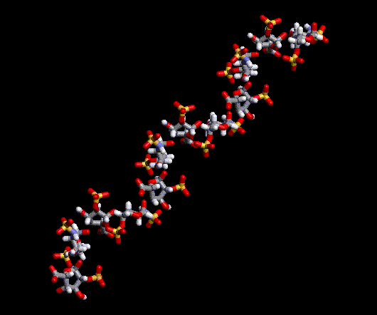
HS/heparin structure
HS and heparin are long, linear chains of sugars, composed of repeating
disaccharide units made up of alternating uronic acid (glucuronic or iduronic
acid) and glucosamine residues. The backbone structure is then decorated
with complex patterns of sulphate groups at various positions.
HS and heparin are long, linear chains of sugars, composed of repeating
disaccharide units made up of alternating uronic acid (glucuronic or iduronic
acid) and glucosamine residues. The backbone structure is then decorated
with complex patterns of sulphate groups at various positions.
For more detailed information concerning the Perichondrium, chondrocytes, PTHrP, Ihh and other signaling
pathways affected by the defect in the EXT genes please view the video link below.
To view this video presentation given by Dr. Henry Kronenberg during this conference please click the link tab
The MHE Research would like to thank all for the use of the presentation of The Perichondrium in Bone Development
on the MHE Research Foundation website. This presentation was from the April 25–28, 2007, the 2nd Conference
on Skeletal Biology and Medicine held in NYC.
This meeting, was jointly hosted at the New York Academy of Sciences and Mount Sinai School of Medicine, was organized and
chaired by Mone Zaidi, professor of endocrinology, geriatrics and adult development, and structural and chemical biology at
Mount Sinai. Cochairs were Gerard Karsenty of Columbia University and Steven Teitelbaum of the Washington University School
of Medicine.
Henry H. Roehl, Ph.D.
Roehl Lab website page
Department of Biomedical Science, The University of Sheffield, United Kingdom
The Roehl laboratory focuses on the role of heparin sulphate proteoglycans (HSPGs) during development of the zebrafish.
Although HSPGs are ubiquitous structural components of the extracellular matrix, they are also thought to play very specific
roles in cell-cell signalling during development. The disaccharide repeats that make up the heparan chains come in 32 different
varieties making heparan sulphate the most information-dense biopolymer found in nature. Binding studies and X-ray
crystallography have identified many specific interactions between oligosaccharides and secreted proteins. Mutational analysis of
genes involved with proteoglycan synthesis has shown that Wingless, Decapentaplegic, Fibroblast Growth Factor and
Hedgehog signalling pathways all depend on proteoglycans at different times during Drosophila development. These data
together have led to the hypothesis that different HSPGs have highly specialized roles including limiting or facilitating signal
diffusion, blocking signal degradation and modulating signal/receptor complex formation.
Our work in this field began with the positional cloning of a small family of zebrafish mutants that all have similar phenotypes
suggesting that their gene products interact or are in the same pathway. These genes, pinscher (pic/papst1), boxer
(box/extl3) and dackel(dak/ext2), are all required for development of the pectoral fins, sorting of the retinotectal projections
and morphogenesis of the skeleton. This cloning project has been a collaboration between three groups (Chi-Bin Chien, U. of
Utah; Robert Geisler, M.P.I. Tuebingen; Henry Roehl, U. of Sheffield). Together, we have found that box and dak encode
glycosyltransferases responsible for synthesis of the heparin sugar chain (EXTL3 and EXT2 respectively), and pic encodes a
sulphur transporter that is involved with the sulphation of all proteogycans (PAPST1). These finding have allowed us to begin
to address the functional requirements of HSPGs during zebrafish development.
Recently we have turned our attention to the role of HSPGs play in a disease called Hereditary Multiple Exostoses (HME). HME is
an autosomal dominant disorder that affects 1 in 50,000 among the general population. Patients with HME have a short stature
and develop numerous cartilage-capped tumours (called exostoses or osteochondromas) from the growth plates of their
longbones. Mutations in human EXT2 account for a large percentage of the cases of this disease. While osteochondromas are
normally benign, they can lead to complications and patients have a 1-2% risk of developing chondrosarcoma or osteosarcoma.
The dominant and sporadic nature of tumour formation in HME patients has led to the proposal of two genetic models.
Osteochondromas may arise from a loss of heterozygosity (LOH) at one of the EXT loci in a developing chondrocyte resulting in
unregulated growth and clonal expansion. In support of this model, somatic mutations or aneuploidy have been found in 3 out
of 46 osteochondromas analysed. The alternative model is that reduced EXT gene dose results in a structural change that
allows chondrocytes to occasionally escape normal developmental constraints to give rise to an osteochondroma.
Please note more research publications can be located on researchers foundation website pages
| ||||||||||||||||||||||||||||||||||||||||||||||||||||||||||||||||||||||||||||||||||||||||||||||||||||||||||||||||||||||||||||||||||

| ||||||||||
Maurizio Pacifici, Ph.D.
Director of the Translational Research Program in Pediatric Orthopaedics website page and Director of
Research, Department of Orthopedic Surgery The Children's Hospital of Philadelphia
The mechanisms by which exostoses form along the growth plates of long bones and other skeletal elements remain largely
unclear. Since the majority of HME patients carry loss-of-function mutations in Ext1 or Ext2, other groups previously created
heterozygous null Ext1 (Ext1+/-) mice that were expected to display traits of HME patients.
Surprisingly, the mutant mice did not completely mimic the human phenotype. Exostosis-like masses were observed in 10-20%
of the mice only, and the ectopic masses were rather small and atypical in organization and were limited to the ribs. Based on
immunohistochemical evidence that human exostoses contain far less heparan sulfate (HS) than would be expected of a
heterozygous Ext mutation (about 50% of control levels), we reasoned that mice producing lower amounts of HS chains may be
able to mimic the human condition more closely. Thus, we created and examined double heterozygous Ext1+/-/ Ext2+/- mice.
Indeed, the double hets mice did display stereotypic exostoses along their long bones that were characterized by a distal
cartilaginous cap followed by a pseudo growth plate and were oriented at a 90 degree angle with respect to the long axis of the
long bones.
We even observed osteochondromas masses at other locations. The data strongly indicate that exostosis formation and
organization are intimately sensitive to, and dependent on, HS production and/or content and that frequency of exostosis
formation can be increased by progressive decreases in Ext expression. Data from an additional mouse model of HME and data
from mesenchymal cell cultures that provide important insights into the mechanisms of exostosis induction and formation. Oub
labs on going work is focused on developing therapeutic approaches for treating the disease.

|
Written consent must be obtained to attach web pages or the files attached to this website, please email the webmaster. Email the webmaster: webmaster@mheresearchfoundation.org Materials on this website are protected by copyright Copyright © 2014 http://eskolab.ucsd.edu/index.shtmlThe MHE Research Foundation Disclaimer: While many find the information useful, it is in no way a substitute for professional medical care. The information provided here is for educational and informational purposes only. This website does not engage in the practice of medicine. In all cases we recommend that you consult your own physician regarding any course of treatment or medicine. This web page was updated last on 3/6/14, 12:0O pm Eastern time |
The MHE Research Foundation, we comply with the HONcode standard for health trust worthy information: By the Health On the Net Foundation.
Click here to verify.# HON Conduct 282463 and is the patient support link on the US Government Genetics Home Reference (http://ghr.nlm.nih.gov)
website, also linked for Patient Information on The Diseases Database a cross-referenced index of human disease, as well as the
Intute: health & life sciences a free online service providing access to the very best Web resources for education and research located in the UK
The MHE Research Foundation is proud to be working with the EuroBoNeT consortium, a European Commission granted Network of Excellence for
studying the pathology and genetics of bone tumors.
studying the pathology and genetics of bone tumors.
| This website is regularly reviewed by members of the Scientific and Medical Advisory Board of the MHE Research Foundation. All online submission forms use (SSL AES 256 bit encryption (High); RSA 1024 bit exchange) Protocol with Privacy protection. Our goal is to make this website as safe and user friendly as possible. |


| The MHE Research Foundation is a participating member organization of the United States Bone and Joint Decade/Initiative, (USBJD/I) & the USBJD/I Rare Bone Disease Patient Network |
The MHE Research Foundation is proud to be an affiliate of the Society For Glycobiology


Please review the publication and video webpages